Friday, 15 February 2013

Unwanted hair in our body

Unwanted hair in our body

The normal amount of body hair for women varies. Most of the time, a woman only has fine hair, or peach fuzz, above the lips and on the chin, chest, abdomen, or back. If you have coarse, dark hairs in these areas, the condition is called hirsutism. Such hair growth is more typical of men.

Causes

Women normally produce low levels of male hormones (androgens). Body makes too much of this hormone, may have the chance to getting unwanted hair growth.
In most cases,hair growth is tends to acumulate depening on the families. In general, hirsutism is a harmless condition.
A common cause of hirsutism is polycystic ovarian syndrome (PCOS). Women with PCOS and other hormone conditions that cause unwanted hair growth and also have acne, problems with menstrual periods, trouble losing weight, and diabetes. If these symptoms start suddenly it getting a chance to, have a tumor that releases male hormones.

Other, rare causes of unwanted hair growth may include:

  • Tumor or cancer of the adrenal gland
  • Tumor or cancer of the ovary
  • Cushing syndrome
  • Congenital adrenal hyperplasia
  • Hyperthecosis (a condition in which the ovaries produce too much male hormones)
  • Use of certain medicines, including testosterone, danazol, anabolic steroids, glucocorticoids.
Rarely a woman with hirsutism will have normal levels of male hormones, and the specific cause of the unwanted hair growth cannot be identified.
ovarian cyst
over productive ovaries

Home Care

Hirsutism is generally a long-term problem. There are a number of ways to remove or treat unwanted hair. Some treatment effects last longer than others.
  • Weight loss in overweight women can reduce hair growth.
  • Bleaching or lightening hair may make it less noticeable.

Temporary hair removal options include:

  • Shaving does not cause more hair to grow, but the hair may look thicker.
  • Plucking and waxing are fairly safe and are not expensive. However, they can be painful and there is a risk for scarring, swelling, and skin darkening.
  • Chemicals may be used, but most have a bad odor.

Permanent hair removal options include:

Electrolysis uses electrical current to permanently damage individual hair follicles so they do not grow back. This This method is expensive, and multiple treatments are needed. Swelling, scarring, and redness of the skin may occur.
Laser hair removal uses laser aimed at the dark color (melanin) in the hairs. This method is best if a very large area needs to be treated and only if the hair is particularly dark (does not work on blond or red hair).

 Contact a Medical Professional

Call your health care provider if:
  • The hair grows rapidly
  • You also have male features such as acne, deepening voice, increased muscle mass, and decreased breast size
  • You are concerned that medication may be worsening unwanted hair growth

Blood tests may be done, including:

  • Testosterone
  • Dihydroepiandrosterone sulfate (DHEA-S)
  • Luteinizing hormone (LH)
  • Follicle stimulating hormone (FSH)
  • Prolactin
  • 17-hydroxyprogesterone
If a tumor is suspected, x-ray tests such as a CT scan or ultrasound may be recommended.

Medications or other treatments.

  • Birth control pills. It may take several months to begin noticing a difference.
  • Anti-androgen medications such as spironolactone may be tried if birth control pills do not work. There is a risk of birth defects if you become pregnant while taking these medicines.
  • Laser hair removal or electrolysis.

Way to remove trouble

There are also some additives you can use in your massage oil that will help to reduce hair growth on the body. Since you have not stated where the hair growth is, general information can be given about it. Make a pasty mix of gram flour, yogurt and a few drops of olive oil. This solution is to be rubbed all over the part of the body that has unwanted hair. This will make the hair come out quite easily. You can apply this paste and leave it on till it dries a bit. Then start rubbing it off. This is the most highly effective method to get rid of unwanted hair. If you repeat this procedure once a week for a few months, you will notice that the hair will gradually stop reappearing. Alternately, you can also try this method. Make a ball of well kneaded dough. Dip this in oil and keep rubbing over the areas where you have hair. This will also reduce hair growth in the area to a very large extent. You need to keep in mind that these practices will have to be followed on a regular basis for results to show up.


Thursday, 14 February 2013

How remedies for acne

Home remedies  for  Acne

  1. Take a few pieces of jaiphal (nutmeg), mix it with some unboiled milk and apply the paste on your acne. Wash it off after about two hours.
  2. - Make a mixture of cinnamon powder and honey and apply on your pimples before you go to bed in the night. Wash it with warm water when you wake up in the morning. Do this for a fortnight and watch your pimples disappear.
  3. - Not only are oranges good for overall health, you can use orange peels as a remedy for pimples too. Grind the peels, mix them with a few drops of water and apply it on your face.
  4. - Mix a tablespoon each of fresh lime juice and groundnut oil and apply it on your face to avoid blackheads.
  5. - Leave fresh mint juice on your face overnight to tame pimples.
  6. - Take methi (fenugreek) leaves and make a paste. Apply it every night for 15 minutes and wash it off.
  7. - Take turmeric powder and neem leaves. Apply the mixture on your pimples. Leave it on your face for about half an hour.
  8. - Take equal quantities of rose water and lime juice and apply the mixture on the affected area. Leave it on for 20 minutes.
  9. - Make a face-wash by adding fresh lime juice to a cup of unboiled milk and using it on your face.
  10. - Take some tomato pulp and apply it on your face. Leave it for 45 minutes and wash it off.
  11. - Take a few drops of gulab jal (rose-water) and mix it with sandalwood. Apply the paste on your face and wash it off after 20 minutes.

Simple pimple home remedies for Acne

  1. Make a tiny clay mask with water add lavendor or tea tree oil for a boost.Leave it for over night.
  2. Swab a layer of apple cider venegar over an offencive  blumish .Dont use repeatly.
  3. Dab a bit of lemon juice apply the skin.do not go out with lemon juice on the skin.lemon makes a photosensitive to skin, it cause some damage.
  4. Mustard powder with honey is apply to skin.Leave it for 15 min.Mustard have high salysilic acid.
  5. Apply fresh garlic. It kills bacteria fast.
  6. Freeze strong green tea into ice cubes.Rub over major blummish.it reduce inflamation and provides antimicrobial action.
  7. Tab twice a day with fresh tomato juice.

Wednesday, 13 February 2013

Pimples

Pimples


A pimple or spot is a kind of acne..Excess oil getting trapped in the pores in the skin .Due to this reason pimples are mainly formed . Some of the varieties are pustules or papules .Pimples can be treated by various acne medications prescribed by a dermatologist.

Blockage of pores.

Sebaceous glands inside the pore which produce sebum. When the outer layers of skin shed , the dead skin cells left behind it become 'glued' together by the sebum. This causes the blockage in the pore, especially when the skin becomes thicker .The sebaceous glands produce more sebum which builds up behind the blockage, and this sebum allow the bacteria to cause pimples including the species Propionibacterium acnes.

Treatment

Over-the-counter medications

Common -counter medications for pimples are benzoyl peroxide and salicylic acid and antibacterial agents such as triclosan. Both medications can be found in many creams and gels used to treat acne (acne vulgaris) through application. Both medications help skin slough off more easily, which helps to remove bacteria faster. Before applying them the patient needs to wash the face with warm water and dry. A cleanser may also be used for that purpose. Acne rosacea is not caused by bacterial infection. It is commonly treated with tretinoin. The most common product is a topical treatment of benzoyl peroxide, which has minimal risk apart from minor skin irritation that may present mild allergy. Recently nicotinamide, is more effective than the antibiotics such as clindamycin. Nicotinamide (vitamin B3) is not an antibiotic and has no side-effects typically associated with antibiotics. It has the added advantage of reducing skin hyperpigmentation which results in pimple scars

Prescription medication

Severe acne usually indicates the necessity of prescription medication to treat the pimples. Prescription medications used to treat acne and pimples include isotretinoin, which is a retinoid. Commonly antibiotics such as tetracyclines and erythromycin were prescribed. While they were more effective than topical applications of benzoyl peroxide, the bacteria eventually grew resistant to the antibiotics and the treatments became less effective. Also, antibiotics had more side effects than topical applications, such as stomach cramps and severe discoloration of teeth.

Hygiene

Practicing good hygiene, including regularly washing skin areas with natural elemens can reduce the amount of dead skin cells and other external contaminants on the skin that lead to the development of pimples.It is not possible to completely prevent pimples , even with good hygiene practices.


How to control fat

 What is fat?

A fat  is a natural oily or greasy substance occurring in animal bodies, esp. when deposited as a layer under the skin or around certain organs.Human being is not  live without  fat.Fats  are  classified as many types which is good or bad.

Factors involved to control fat  

Eat breakfast

  • Try to eat breakfast at the same time each day. Eat breakfast as we waking up beause it keeps the insulin level steady and LDL level is low.(bad cholesterol.)
  • Protein and high-fiber foods are taking the breakfast — because they take longe t ime to digest than refined sugars and complex carbs, we feel continue throughout the morning. Try eggs, peanut butter, and fresh fruit and vegetables.
  • Avoid making sugary cereals, waffles, pancakes, french toast, breakfast pastries, or instant oatmeal the sole focus of your breakfast. While we take this type of food we are try to balance with protein or fiber 
    Decompress. Research indicates that the secretion of cortisol (a hormone your body produces1 during times of stress) is correlated with an increase in belly fat. . Here are some tips for combating everyday stress:
  • Get enough sleep. Most adults need at least 7 hours of sleep every night to function properly.
  • Set time to relax. Even if it's only 15 minutes on your lunch break, try to find time to simply close your eyes, breathe deeply, and forget your worries.
  • Keep your stressors away from your sleeping area. If you can avoid it, don't do work or anything stressful in your bedroom. Tell yourself that the area is reserved for rest and relaxation, and resolve to leave your worries behind as soon as you step into the room.
Aim to take 10,000 steps a day.
  • Walking is a good exercise for reducing fat.Keep walking everyday in our life.
  • Get a pedometer and try to increase the number of steps you take each day.
  • Take the stairs instead of the elevator. Walk instead of driving.
  • Stand up and walk for 30 steps every 30 minutes.
Switch out refined grains
 
People taking low fat diary,fish, meat, fruits and vegetables are balance quantity.It reduce the fat belly.
Melt fat with grains. 
A diet rich in whole grains changes the glucose and insulin response in your body, which avoid melting of fat, and visceral fat, that deep layer of fat, is easier for your body to burn than the subcutaneous fat under your skin.Try to avoid "white" grains. Instead of brown wheat bread is used 
  • Drink plenty of water 
    . Studies suggest that consistently drinking water throughout the day can lead to a more active metabolism, Additionally, drinking more water helps your body flush out waste and toxins and improves your overall health.
    • Aim to drink an 8-10 glass of water 8 times per day, or 64 ounces total.
    • Carry a water bottle with you so that you can drink whenever you feel thirsty.
    • Know how to tell when you're sufficiently hydrated. It's a slightly gross metric, but you'll know you're drinking enough water when your urine runs almost clear. If it's still yellow, drink up.

      Two: Exercising for Fat Loss 

      Exercise in small bursts

. Run fast and as far as you can for 20 seconds, then slow to a walk until you catch your breath. Repeat for 10 minutes.
  • Set a treadmill, elliptical or stationary bike for interval training. Most modern exercise equipment can be set to an interval training mode, which will significantly increase the difficulty of the exercise for short periods of time.
  • Go for a quick walk. Take long strides and keep a brisk pace, or try going up and down a flight of stairs.

Skip the crunches — for now.

  • Doing abdominal crunches and sit-ups will build strong muscles, but you might not be able to see them under the remaining belly fat.
  • Do squats. Stand with your feet 8-9 inches apart, extend your arms in front of you, and squat your hips backward. Do four sets of 15 to 20 squats, working several minutes at a time.
  • Stretch the sides of your waist. Stand up straight, with your feet hip-width apart. Put your right hand on your right hip, and lift your left arm straight up, so that your palm is facing to the right. Keeping your legs centered, lean to the right and "reach" over with your left arm, stretching your left side. Do 3 to 5 times on each side.
Ramp up the cardio. 
Aerobic exercises, which get your heart pumping, burn calories quickly and will facilitate fat-loss all over your body, including your belly.
  • Time your miles.  

    Track your progress by timing how long it takes to run a mile. As our cardiovascular stamina improves, we will notice the time going down.

  • Dont' overdo it. 
    1. When you're first getting into cardio, aim to workout 3 days per week, then build up to 4 when you're ready. Pushing yourself hard every single day won't give your body enough time to recover and build up muscle, and could lead to injury .
    2. aeficit to lose one pound of fat. That is, you have to either burn off 3700 calories through exercise or eat 3700 calories less than you burn in a week..
    3. Keep a food diary. Most people tend to underestimate how much they eat in a day.
    4. Try a diet in which you consume 2200 calories (men) or 2000 calories (women) per day. This should cause a deficit sufficient for you to lose one or two pounds per week, depending on your activity level.

    Eat good fats.

    1. Studies suggest that a diet with a higher ratio of monounsaturated fats (MUFAs) — such as avocados, nuts, seeds, soybeans, and chocolate — can prevent the accumulation of belly fat.

    2. Trans fats (in margarines, crackers, cookies, or anything made with partially hydrogenated oils) seem to result in more fat deposited in the abdomen, so avoid these as much as you can.
    3. Get more fiber in your diet. Soluble fiber (such as that found in apples, oats, and cherries) lowers insulin levels, which, as mentioned earlier, can speed up the burning of visceral belly fat.
    4. Add fiber to your diet slowly. If you are currently getting 10 grams of fiber a day, don't jump to 35 grams of fiber the next day. You need to give the natural bacteria in your digestive system time to adapt to your new fiber intake
    5. Leave the skin on your fruits and vegetables. Incorporating more fruits and vegetables into your diet will add fiber, but only if you eat the skin, because that's where all the fiber is. So don't peel those apples before you eat them. If you're eating potatoes, try to leave the skin in the dish (such as if making baked or mashed potatoes) or if you peel them, make a snack out of them, such as baked garlic Parmesan peels. It's also worth knowing that keeping the skin on potatoes when you cook them will help keep more vitamins and minerals in the flesh.
    6. Eat more split pea soup. Split peas is a fiber "power food". Just one cup of them contains 16.3 grams of protein.

Tuesday, 12 February 2013

Okra

 Main  benefits  of Okra


1) The main fiber content in Okra helps to stabilize blood sugar by conform the rate at which sugar is absorbed from the intestinal tract.
2) Okra's mucilage binds cholesterol and bile acid which carrying toxins dumped into and it filter by liver.
3) Okra helps lubricate the large intestines due to its bulk laxative qualities. The Okra fiber absorbs water and ensures bulk in stools. This helps improve constipation. , Okra's mucilage content is act as main role. Okra elimination more comfortably by its slippery characteristic. Okra help to passing the waste easily from our body. Okra is completely non-toxic, non-habit forming, has no adverse side effects, is full of nutrients, and is economically within reach of most unlike the OTC drugs.
4) Okra fiber is excellent for feeding the good bacteria (probiotics). This contributes to the health of the intestinal tract. This helps to biosynthesis of Vitamin B complex.
5) Okra is good for summer heat treatment. Okra is a supreme vegetable for those feeling weak, exhausted, and suffering from depression.
6) Okra is used for healing ulcers and to keep joints limber. It helps to neutralize acids, being very alkaline, and provides a temporary protective coating for the digestive tract.
7) Okra treats lung inflammation, sore throat, and irritable bowel.
8)  Okra has been used successfully in experimental blood plasma replacements. To retain most of Okra's nutrients and self-digesting enzymes, it should be cooked as little as possible, e.g. with low heat or lightly steamed. Some eat it raw.

Other benefits

  • Okra is used to reduce weight and it is cooked over low flame to retain its properties. This also to ensure that the invaluable mucilage contained in it is not lost to high heat.
    For adding bounce your hair, boil horizontally sliced Okra till the brew become maximally slimy. Cool it and add a few droops of lemon and use this as the last rinse and see your hair spring back to youthfulness and jump.
  • Okra is an excellent laxative treats irritable bowels, heals ulcers and problem in the gastrointestinal track.
  • Protein and oil contained in the seeds of Okra serves as the source of first-rate vegetable protein. It is enriched with amino acids such as tryptophan, cystine and other sulfur amino acids.
  • Vitamin C is a powerful antioxidant and anti-inflammatory. This anti-inflammatory agent cut the development of asthma symptoms. Vitamin C rich fruits are have the power to control asthma. 1/2 cup of cooked Okra contains over 13 mg of vitamin C.
  • Okra, are assoc with protection against heart disease in both men and women.
  • The insoluble fiber found in Okra helps to keep the intestinal tract healthy, decreasing the risk of some forms of cancer, especially colon-rectal cancer.
  • Eating plenty of flavonoid and vitamin C-rich fruits and vegetables such as Okra helps to support the structure of capillaries.
  • 1/2 cup of cooked Okra contains 460 IU of vitamin A. eating more foods rich in beta-carotene or vitamin A was associated with a lower risk of cataracts.
  • . The diet was rich in soluble fiber from oats, barley, psyllium, eggplant and Okra. It used soy substitutes instead of meat and milk and included almonds and cholesterol-lowering margarine every day.

Dandruff

Dandruff  definition

Dandruff is common scalp disorder is refers to the shedding of dead skin cells from the scalp. Dandruff should not be confused with a simple dry scalp. Most cases of dandruff can be easily treated with specialised shampoos. There is, however, no true cure. It affect half of the population.

Causes

As the epidermal layer replaces itself, cells are pushed outward where they eventually die and flake off. For most individuals, these flakes of skin are too small to be visible. certain conditions cells are goes to unusual rapid especially in the scalp. For people with dandruff, skin cells may mature and be shed in 2–7 days, as opposed to around a month in people without dandruff. The result is that dead skin cells are shed in large, oily clumps, which appear as white or grayish patches on the scalp, skin and clothes.
Dandruff is formed due to these factors,
  1. Skin oil commonly referred to as sebum or sebaceous secretions.
  2. The metabolic by-products of skin micro-organisms (most specifically Malassezia yeasts)
  3. Individual susceptibility
In older days fungus Malassezia furfur as the cause of dandruff. While this species does occur naturally on the skin surface of both healthy people and those with dandruff. Later the specific fungus is responsble for scalp named as, Malassezia globosa, It metabolizes triglycerides present in sebum by the expression of lipase, resulting in a lipid byproduct oleic acid (OA). During dandruff, the levels of Malassezia increase by 1.5 to 2 times its normal level.

Treatment

Shampoos use a combination of ingredients to control dandruff. The pathogenesis of dandruff involves hyper proliferation of keratin cites, resulting in deregulation of keratinization. The corneocytes clump together, formed as large flakes of skin. Essentially, keratolytic agents such as salicylic acid and sulphur loosen the attachments between the corneocytes and allow them to get swiped off.

Regulators of keratinization

Zinc pyrithione (ZPT) heals the scalp by regulating the epithelial keratinization or sebum production or both.Significant reduction in the number of yeasts after use of ZPT, which is an antifungal and antibacterial agent The population of Malassezia decreases, parakeratosis gets eliminated and corneocytes lipid inclusions are diminished.

Steroids

The parakeratotic properties of topical corticosteroids depend on the structure of the agent, the vehicle and the skin onto which it is used. Corticosteroids work via their anti-inflammatory and antiproliferative effects

Selenium sulfide

It is believed that selenium sulfide controls dandruff .Its anti Malassezia effect rather than by its antiproliferative effect, although it has an effect in reducing cell turnover. It has anti-seborrheic
properties as well as cytostatic effect on cells of the epidermal and follicular epithelium. The excessive oiliness after use of this agent has been reported in many patients as adverse drug effect.

Coal Tar

Coal tar is a keratoplastic agent causing the skin to shed dead cells from the top layer and slow skin cell growth.

Imidazole antifungal agents

Imidazole antifungals such as ketoconazole act by blocking the biosynthesis of ergosterol, the primary sterol derivative of the fungal cell membrane. Changes in membrane caused by ergosterol depletion or incompatible with fungal growth and survival.
Ketoconazole is a broad spectrum, antimycotic agent that is active against both Candida and M. furfur . Of all the imidazoles, ketoconazole has become the leading contender among treatment options because of its effectiveness in treating seborrheic dermatitis as well.

Hydroxypyridones

In contrast to the imidazole antifungals, the hydroxypyridones do not affect sterol biosynthesis; instead they interfere with the active transport of essential macromolecule precursor, cell membrane integrity and the respiratory process of cells. Ciclopirox is widely used as an anti-dandruff agent in most preparations

Neem

Neem has anti-fungal properties that can be effective in treating dandruff.

Black Pepper

Black pepper contains the dandruff fighting minerals zinc and selenium. In Indian traditional Ayurveda, powdered black pepper has been used to treat dandruff.Egg oil is also treat dandruff.

Sandalwood

Sandalwood and Lemon: Application of an emulsion prepared with the oil of sandalwood and lemon juice has been proven effective to cure dandruff. In traditional Ayurveda, sandalwood has many benefits. Its main component beta-santalol (~90%) has antimicrobial properties.


Monday, 11 February 2013

Dengu fever


What is dengue fever?

Dengue (pronounced DEN gee) fever is a painful, debilitating mosquito-borne disease caused by any one of four closely related dengue viruses. These viruses are related to the viruses that cause West Nile infection and yellow fever.Mosquito named as Aides mosquito.It is not spread  to  one  person  to  another  person.
signs of dengue fever include bleeding gums, severe pain behind the eyes, and red palms.

Dengue  can affect anyone but  it more severe in people with compromised immune systems. Because it is caused by one of four stereotype's of virus, it is possible to get dengue fever multiple times.  Attack of dengue produces immunity for a lifetime to that particular stereotype to which the patient was exposed.

Dengue hemorrhagic fever is a more severe form of the viral illness. Symptoms include headache, fever, rash, and evidence of hemorrhage in the body. Petechiae (small red or purple splotches or blisters under the skin), bleeding in the nose or gums, black stools, or easy bruising are all possible signs of hemorrhage. This form of dengue fever can be life-threatening and can progress to the most severe form of the illness, dengue shock syndrome.

Symptoms of Dengue Fever

Symptoms, which usually begin four to six days after infection and last for up to 10 days, may include
  • Sudden, high fever
  • Severe headaches
  • Pain behind the eyes
  • Severe joint and muscle pain
  • Nausea
  • Vomiting
  • Skin rash, which appears three to four days after the onset of fever
  • Mild bleeding (such a nose bleed, bleeding gums, or easy bruising)
Sometimes symptoms are mild and can be mistaken for those of the flu or another viral infection. However, serious problems can develop. These include dengue hemorrhagic fever, a rare complication characterised by high fever, damage to lymph and blood vessels, bleeding from the nose and gums, enlargement of the liver, and failure of the circulatory system. The symptoms may progress to massive bleeding, shock, and death. This is called dengue shock syndrome (DSS)

Diagnosing Dengue Fever

Doctors can diagnose dengue infection with a blood test to check for the virus or antibodies to it. If you become sick after travelling to a tropical area, let your doctor know.This will indicate he patient is infect with dengue.

Treatment for Dengue Fever

Today there is a number of specific medicines are used to treat the dengue . If you think you may have dengue fever, you should use pain relievers with acetaminophen and avoid medicines with aspirin, which could worsen bleeding. You should also rest, drink plenty of fluids, and see your doctor.
If you start to feel worse in the first 24 hours after your fever goes down, you should get to a hospital immediately to be checked for complications.We are awaking to reducing mosquito .The dump of water is settled in the tanks This easily cause the infection.

Jaundice

 Jaundice


Jaundice, is a term used to describe a yellowish tinge to the skin ,mucus membrane,and sclerae (the white part of the eye) that is caused by hyperbilirubinemia that means an excess of bilirubin in the blood resulting increase level of plasma. Body fluids may also be yellow. The color of the skin and sclerae varies depending on levels of bilirubin; mildly elevated levels show yellow skin and sclerae, highly elevated levels shows brown.
Bilirubin is a waste product that remains in the bloodstream after the iron is removed from the hemoglobin.It is done by the process called degradation of erythrocytes (cells that contain hemoglobin and can carry oxygen to the body). Excess of bilirubin is passed to the surrounding tissues, saturating them with this yellow substance.
Bilirubin is circulating freely in the blood is called unconjugated bilirubin. Once it present in the liver, other chemicals are combined with bilirubin, creating a substance called conjugated bilirubin, is formed in the bile and then excreted. Bilirubin is what gives feces its brown color.

Types of jaundice:

  • Hepatocellular jaundice - a type of jaundice that occurs as a result of liver disease or injury.
  • Hemolytic jaundice - a type of jaundice that occurs as a result of hemolysis (an accelerated breakdown of erythrocytes - red blood cells) leading to an increase in production of bilirubin.
  • Obstructive jaundice - a type of jaundice that occurs as a result of an obstruction in the bile duct (a system of tubes that carries bile from the liver to the gallbladder and small intestine), which prevents bilirubin from leaving the liver.

 causes of jaundice

These are the matters are mostly cause jaundice. They are,
  • Acute inflammation of the liver - may impair the ability of the liver to conjugate and secrete bilirubin, resulting in a buildup of bilirubin.
  • Inflammation of the bile duct - may prevent the secretion of bile and removal of bilirubin, causing jaundice.
  • Obstruction of the bile duct - prevents the liver from disposing of bilirubin, which results in hyperbilirubinemia.
  • Hemolytic anemia - Production of bilirubin increases when large quantities of erythrocytes are broken down.
  • Cholestasis-a condition in which the flow of bile from the liver is interrupted. The bile containing conjugated bilirubin remains in the liver instead of being excreted.

symptoms of jaundice

Most common symptoms is yellowish in skin,and sclerae.Some of other symptoms includ
  • Pruritis (itchiness)
  • Fatigue
  • Abdominal pain - typically indicates a blockage of the bile duct.
  • Weight loss
  • Vomiting
  • Fever
  • Paler than usual stools
  • Dark urine

Diagnosis of jaundice

Diagnosis of jaundice is depends upon the attention of abdomen. Doctors will be feeling for masses (tumors) in the abdomen and checking the firmnes of the liver; a firm liver indicates cirrhosis, while a rock-hard liver indicates cancer.

The severity of jaundice is determined by liver function test.
If the cause of your symptoms cannot be identified, your doctor may require blood tests to check levels of bilirubin and evaluate the composition of the blood. Some of these tests include:
  • Bilirubin tests - a high level of unconjugated bilirubin relative to levels of conjugated bilirubin indicate hemolysis (accelerated break down of erythrocytes) 

  • Full blood count (FBC), or complete blood count (CBC) - measures levels of erythrocytes (red blood cells), leukocytes (white blood cells), and thrombocytes (platelets).
  • Hepatitis A, B, and C tests
If an obstruction of the liver is suspected, the liver's structure will be looked at with the help of imaging tests. Some of these tests include:
  • Magnetic resonance imaging (MRI) - uses magnetic signals to create image "slices" of the soft tissues of the human body. MRIs can also be manipulated in such a way that the cause and location of a bile duct obstruction can be identified.
  • Abdominal ultrasonography (ultrasound) - uses high frequency sound waves to create a two-dimensional image of the soft tissues inside the human body. It is especially useful in identifying gallstones, although tumors and dilated bile ducts may also show.
  • Computerized tomography (CT) scan, or computerized axial tomography (CAT) scan - uses a thin X-ray beam to create image "slices" of soft tissues in the body. It is especially useful in identifying tumors and dilated bile ducts.
A liver biopsy is particularly useful in checking for inflammation, cirrhosis, cancer, and fatty liver. This test involves injecting a needle through the skin and into the liver to obtain a sample of the tissue, which is examined under a microscope.

 Treatment for jaundice 

Anemia-induced jaundice may be treated by increasing the amount of iron in the blood; either by taking iron-supplements or eating more iron-rich foods.
Hepatitis-induced jaundice may be treated with anti-viral or steroid medications.
Obstruction-induced jaundice may be treated via surgery to remove the obstruction.
Medication-induced jaundice is treated by selecting an alternative medication and by discontinuing medications that caused jaundice.

Prevention of jaundice

Jaundice is related to the function of the liver, so it is essential that you maintain this vital organ's health by eating a balanced diet, exercising at least 30 minutes five times a week, and refraining from exceeding recommended amounts of alcohol.


Wednesday, 6 February 2013

Kidney stone treatment part 2

 Laser treatment for kidney stone


   Laser lithotripsy isan advace treatment can be performed under general anesthesia, This procedure can be used to remove or break up (fragment) stones located in the ureter or kidney. A special telescopic instrument ccalled, flexible ureteroscope is inserted through the urethra and passed through the bladder and up the ureter to the stone. The light energy of the laser is transported through a light guide to the stone. The important factor is fiber is very thin it can bend with the flexible ureteroscope to reach any location of the kidney .
Stone fragmentation occurs when pulses of intense laser light are applied. The fragments are then passed by the patient or removed with a basket.

Advantages of Laser Kidney Stone Treatment

 
  •  We dont need open surgery
  • We can identify stones any location in ureter or kidney with 99% success rate.
  • We can treat stones up to 3 cm.
  • One day of hospital stay only.
  • We can back to normal duties with in 4 days.
  • Best option in bleeding diathesis.
  • Best option for hard stones.
  • Best option for stones in very dependent lower calyx.
  • Best option for post PCNL residual stones.
  • Best treatment for Pilots with multiple small kidney stones.
  • Best treatment for Pregnant women, morbid obesity,

Keyhole surgery

Keyhole Surgery (PCNL) is a well-established procedure by which stones in the kidney or the upper ureter are removed by making a small incision in the flank. Generally, an incision, that is 1 cm or less than 1 cm, is made in the flank. A guide wire is passed through this incision into the kidney. Keyhole Surgery is performed under fluoroscopy or x-ray control.

Method :

A passage is then created around this guide wire by dilatation. Through this passage, a nephroscope is passed into the kidney to visualise the stone and remove it. Larger stones can be fragmented by different methods and removed. Stones are therefore cleared easily. Once the procedure is complete, a tube is left through this tract as drainage for one or two days.

Advantage & Other notes:

The main advantage of this method is need not open surgery, only a 1 cm incision is made in the flank. The stones can be visualised directly and removed. Unlike ESWL or ureteroscopy, the stones are removed in the same sitting and the kidney is cleared of calculi. The stay in the hospital is only for 3-4 day

This surgery would be recommended as a treatment of choice, if the patient has kidney stones larger than 2 cm

This procedure is commonly performed under general anaesthesia, and therefore you also need to be admitted to the hospital for this procedure.

The success rate of clearance of stones with this procedure ranges between 90 to 95%. This actually depends upon the size, number and location of these stones. Sometimes, complete clearance may require a second procedure after a few days.

For the post-operative course, the patient will generally need to stay in the hospital for 2 to 3 days after the procedure. Patient examine the studies if any stone is prsent in the kiney.

Rare complications include persistent uncontrolled bleeding due to arteriovenous malformations or pseudo-aneurysms (0.4%), which would need secondary procedures. When a supracostal puncture (above the 12th rib) is made, complications would then include pneumothorax (commonly called collapsed lung caused due to the accumulation of air or gas in the space surrounding the lungs) or fluid accumulation in the thorax. Again, the incidence of these is only 0-4%.

Most patients return to average activity levels within a week or so. A return to vigorous activity should probably take place after 4 weeks.

Link here for Kidney stone part 1


Kidney stone part 1

 Kidney stones


Kidney stones usually comprised of a compound called calcium oxalate, are the result of an accumulation of dissolved minerals on the inner lining of the kidneys. These deposits can grow to the size of a golf ball while maintaining a sharp, crystalline structure.
Kidney stones that remain inside the body can lead to many conditions, including severe pain and ureter (the tube connecting the kidney and bladder) blockage that obstructs the path urine uses to leave the body.

 causes of kidney stones

The main reason of kidney stones is a lack of water. Every indiviual should drink eight to ten glass of waer per day. In sufficient water is reae he problem in the result of rising the pH level in the kidney is drop and become acidic. An excessively acidic environment in the kidneys is conducive to the formation of kidney stones.

Medical conditions such as renal tubular acidosis, hyperparathyroidism, medullary sponge kidney, and Dent's disease have been known to lead to kidney stones. It also has been suggested that water fluoridation - the addition of fluoride to drinking water - is responsible for some cases of kidney stones.
Certain medications can increase the risks of developing kidney stones. Scientists found that opiramate (Topamax), a drug commonly prescribed to treat seizures and migraine headaches, can increase the propensity of calcium phosphate kidney stone. Overweight is ormed he uric acid stones.

High in protein and sodium but low in calcium content food, obesity, high blood pressure, and conditions that affect how calcium is absorbed in the body such as gastric bypass surgery, inflammatory bowel disease, and chronic diarrhea.

symptoms of kidney stones

A kidney stone usually remains symptomless until it moves into the ureter.
  • Severe pain in the groin and/or side
  • Blood in urine
  • Vomiting and nausea
  • White blood cells or pus in the urine
  • Reduced amount of urine excreted
  • Burning sensation during urination
  • Persistent urge to urinate
  • Fever and chills if there is an infection

Diagnosis

A physical examination of the kidney stones is colicky pain (in the groin) and pain in the lower back by the kidneys. An analysis of the urine will indicate whether or not there is blood in the urine and if there is a subsequent infection. Blood tests can be done to identify complications that may accompany a kidney stone and check the validity of the diagnosis.

A CT scan of the abdomen is the most thorough way to test for kidney stones. A CT scan will conform the state of the ureter, bladder, and kidneys, whether or not a stone exists, the kidney stone's exact size and location, whether or not a blockage exists, and the state of the other adjacent organs such as the appendix, aorta, and pancreas. Pregnant women may receive an ultrasound rather than a CT scan in order to avoid unnecessary radiation

Prevention

Preventing kidney stones can be very easy as staying hydrated.for beter healh. Diluting the urine with water keeps the minerals from concentrating and forming stones. When one's urine is clear, rather than yellow colored, the chance of stone formation is reduced. Doctors may also prescribe medicines to prevent certain types of stones for individuals who are at higher risk.

Glass of orange juice can help to prevent the recurrence of kidney stones better than other citrus fruit juices such as lemonade.

Link  here for Kidney stone part 2

Tuesday, 5 February 2013

Diabetes

Diabetes

Diabetes, often referred to by doctors as diabetes mellitus, is the person have high blood glucose level either because insulin production is inadequate, or because the body's cells do not respond properly to insulin, or both. Patients with high blood sugar will typically experience polyuria (frequent urination), they will become increasingly thirsty (polydipsia) and hungry (polyphagia).
     Diabetes (diabetes mellitus) is classed as a metabolism disorder. Metabolism refers what we take breakdown into glucose.Glucose is the form of sugar.The sugar is added to the blood stream and give energy to the body .
    When our food is digested, the glucose makes its way into our bloodstream. Our cells use the glucose for energy and growth. However, glucose cannot enter our cells without insulin being present - insulin makes it possible for our cells to take in the glucose.
    Insulin is a hormone that is produced by the pancreas. After eating, the pancreas automatically releases an adequate quantity of insulin to move the glucose present in our blood into the cells, as soon as glucose enters the cells blood-glucose levels drop.A person with diabettes have blood sugar level is too low is called hyperglycemea.

Types of diabetes

Type 1 diabetes
Type 2 diabetes
Gestational diabetes

Type 1 diabetes

In this condition insulin is not produced by pancreas in the patient.Patient should take insulin injection rest of their life and following a heealthy diet.

Type 2 diabetes

In this condition enough insulin not produce by pancreas for regular action.Patient conrol this type by reduce their weight,healthy diet,and check blood glucose level properly.Patient take insulin in the tablet form .Men whose testosterone levels are low have been found to have a higher risk of developing type 2 diabetes.

Gestational diabetes

This type affect women during pregnency. Some women have very high levels of glucose in their blood, and their bodies are unable to produce enough insulin to transport all of the glucose into their cells, resulting in progressively rising levels of glucose .Undiagnosed or uncontrolled gestational diabetes can raise the risk of complications during childbirth. The baby may be bigger than he/she should be.

Diagnosis.

Diabetes is determind by three following tests.They are,
The A1C test
The OGTT (oral glucose tolerance test)
The FPG (fasting plasma glucose) test

Treatment for diabetes

Diabetes type 1 lasts a lifetime, there is no known cure. Type 2 usually lasts a lifetime, however, some people have managed, through a lot of exercise, diet and excellent body weight control to get rid of their symptoms without medication
Patients with type 1 are treated with regular insulin injections, as well as a special diet and exercise.
Patients with Type 2 diabetes are usually treated with tablets, exercise and a special diet, but sometimes insulin injections are also required.

Diabetes Spreads In Southeast Asia

Diabetes is rapidly spreading in Southeast Asia as people embrace American fast foods, such as hamburgers, hot dogs, French fries and pizza. More Chinese adults who live in Singapore are dying of heart disease and developing type 2 diabetes than ever before.

The authors found that Chinese adults in Singapore who eat American-style junk foods twice a week had a 56% greater risk of dying prematurely form heart disease, while their risk of developing type 2 diabetes rose 27%, compared to their counterparts who "never touched the stuff". There was a 80% higher likelihood of dying from coronary heart disease for those eating fast foods four times per week.

Monday, 4 February 2013

strawberry

 Strawberry fruit

      Srawberry are the delicious fruit whih is essencially rich in nutrient for our health.It appears in contrasting color like redish.Mostly it was ocured in hill places like ooty.Ancient roman peoples are believed this fruit is a symbol for attacks,fevers,kidney stones.Strawberries are the only fruit with seeds on the outside.
The average strawberry has 200 seeds.Strawberries are the first fruit to ripen in the spring.
    Strawberries are a member of the rose family.Strawberries are low fat, low calorie; high in vitamin C, fiber, folic acid, potassium.Strawberry is to reduce he risk of cancer,and heart attack.
In medieval times, strawberries were served at important functions to bring peace & prosperity.
Over 53 percent of seven to nine-year-olds picked strawberries as their favorite fruit.
Eating strawberries, which are rich in nitrate, can increase the flow of blood & oxygen to the muscles by 7%.  This prevents muscle fatigue, making exercise easier.
Actually, strawberries are a goitrogen and are bad for those with hypothyroidism. Strawberries are also good source of iodine, for the thyroid, a so frequently over-ignored health problem.


Apple fruit

Apple fruit

Apple fruit is one of the most popular delicious fruit.This wonderful fruit is covered with rich photo-nutrient that is essentially for health. The antioxidants in apple have much health promoting and disease prevention properties.Sscientific name of Apple is Mal us domestica.Shape of the apple is oval and it skin has various colours depending upon cultivation

Health benefits of apple

Apple fruit major role in our body.They contain vitamins ,minerals,anti-oxidants.That functions are given below.
  • Apple fruit contain photo-nutrients, and anti-oxidants. This components are essential for normal growth, development and overall well-being.
  • Apples are low in calories; They, contain no saturated fats or cholesterol. Apple fruit is rich in dietary fibre, which helps prevent absorption of dietary-LLD or bad cholesterol in the gut. The fibre also saves the colon mucous membrane from exposure to toxic substances by binding to cancer-causing chemicals inside the colon.
  • Apples are rich in antioxidant photo-nutrients.The total measured anti-oxidant strength (OR AC value) of 100 g apple fruit is 5900 TE. Some of the important flavonoids in apples are quercetin, epicatechin, and procyanidin B2.They contain tartaric acid that gives tart flavor to them. Altogether, these compounds help the body protect from deleterious effects of free radicals.
  • Apple fruit contains vitamin-C and beta-carotene. Vitamin C is a powerful natural antioxidant. Consumption of foods rich in vitamin C helps the body develop resistance against infectious agents and scavenge harmful, pro-inflammatory free radicals from the body.
  • Further, apple fruit is a good source of B-complex vitamins such as riboflavin, thiamine, and pyridoxine (vitamin B-6). Together these vitamins help as co-factors for enzymes in metabolism as well as in various synthetic functions inside the body.
  • Apple also contains a small amount of minerals like potassium, phosphorus, and calcium. Potassium is an important component of cell and body fluids helps controlling heart rate and blood pressure; thus, counters the bad influences of sodium.

          Selection and Storage

Fresh apples are readily available in the stores all around the season. Choose fresh, bright, firm textured apples with rich flavor. Avoid fruits with pressure marks over their surface as they indicate underlying mottled of pulp.

Fresh apples can be kept at room temperature for few days and stored inside the refrigerator for two to three weeks. Wash them in clean running cold water before use to remove any surface dust and pesticide/fungicide residues.

Cultivation of apple

Tree is Young

One important point in apple cultivation is that they grow best in groups to promote pollination. If you have different varieties, make sure they will all bear pollen at the same time. Apple trees have the best fruit production between the ages of 10 and 30. Most apple trees live to about 35 years of age..
 
When your fruit tree is between 5 and 10 years be sure to:
  • Provide enough air circulation in and around the tree.
  • Prune to establish the essential structure of the tree and then to encourage fruit to develop.
  • Irrigate your tree with a good deep soaking every 7 to 15 days, depending on the season and weather.
  • Check the location of your trees. If they are growing in a lower part of your yard, cold air can accumulate down there, and slow down fruit production.The location is extremely windy/cold/wet during pollination time, this can prevent pollination.

Not Bearing Fruit Yet!

Apple tree is more than 10 years old, still is not bearing fruit, you may want to consider stressing your apple trees. This can be done by cutting back on nutrients and water for a short period of time. You can also stress the tree by taking a straight-edged spade and driving it into the ground in 6-8 places outside the drip-line of the tree. This will cut many of the feeder roots. This type of stressing usually results in blooming and bearing fruit the following year.. Be sure to keep all fertilisers away from the roots.

Pruning Your Apple Trees

Before pruning project check apple trees a good one .Make sure the trunk is not full of rot or large holes. Is most of the wood still alive? If it appears that over half of the branches are dead or splitting apart, the tree may not be worth saving.Bearing apple fruit are check to suitable for cooking,eating and taste it whether it is bitter or sour or sweet.
The trees may be a poor variety if the fruit is small, green, hard and/or sour.
The apple trees are worth pruning, take the time to clean the area around the trees. Clear out any weeds, brush or other non-fruit trees in the area.
Pruning is started from he top into bottom way,this will allow to well ripening fruit. Some people routinely cut off the top third of their apple tree branches to let the sunlight in.. Keep top branches short and the ones below them increasingly long. This allows the tips of all branches to receive light.

Maintenance Pruning

In maintenance pruning is to remove water sprouts as soon as they develop. These sprouts can come up from the branches and also from suckers at the base of the trunk. Water sprouts often develop when the tree is being pruned too much. .
When pruning, make all your cuts back to a live branch or to the trunk. Make your cut just outside the branch collar, so you do not leave a stub. If the branch is long and/or thick, make several cuts before cutting just outside the branch collar. This helps avoid tearing due to the weight and stress on the branch. Remove any limbs that have woodpecker holes. Also cut off branches with weather damage and ones that show signs of disease.

Use Sharp, Clean Tools

Be sure to clean the blade after each cut, and especially before starting to prune a different tree. If your apple trees have been neglected, it’s hard to be sure where diseased wood is. Cleaning the blade will prevent the spread of disease to different branches and trees.
. Be patient, give your trees the proper environment for growing and provide regular pruning to enhance fruit production. Not only will you enjoy the fruit, but apple trees add a lot of character and beauty to your landscape.

Exercises for knee pain part 2

 Knee pain exercises

  1. Wall slide: Leaning with your back against a wall, bend your knees 30°, sliding down the wall, then straighten up again. Move slowly using hands on the wall for balance.Keep feet parralell o not allow knees to go out over the toes
  2. Bent-Leg Raises: Sitting in a chair, straighten one leg in the air (without locking the knee). Hold for about one minute. Bend your knee to lower the leg about halfway to the floor. Hold for 30 seconds. Return to starting position. Work up to 4 reps on each leg.
  3. Straight-Leg Raises: Sitting in a chair, rest your foot on another chair. Lift the foot a few inches off the chair while keeping your leg straight. Hold for 5 -10 seconds. Return to resting position. Repeat 5 -10 times. (Also work on increasing the time, up to 2-3 minutes if possible.)
  4. Abductor Raise: Lie on your side, propped on one elbow. The leg on the floor bent, the other straight. Slowly lift the top leg, hold for 5 -10 seconds, then lower. (Ankle weights will increase the intensity). Do 1-3 sets with 12-15 repetitions each. Remember to rest in between sets.
  5. Hamstring Curl: Stand with the front of your thighs against a surface (a table or wall). Flex one knee up as far as is comfortable. Hold for 5 - 10 seconds, then lower slowly. If possible, do not touch the floor between repetitions. (Ankle weights will increase the intensity.) Do 1-3 sets with 12-15 repetitions each. Remember to rest in between sets.
  6. Step-Ups: Stand in front of a step, like a sturdy bench or stairs, about two feet high (or less if necessary). Step up onto the support, straighten your knees fully (without locking them) and step down. Maintain a steady pace. If you are comfortable with your balance, pump your arms while doing this exercise. Start with 1 minute, slowly building your time. Gets your heart pumping too.

Symptoms of knee pain

  Many symptoms reason for the knee pain.One important symptoms is Swelling

Swelling:

Swelling of the knee is common with several different knee problems. When there is an effusion immediately after a knee injury, a possible cause is severe injury to an internal joint structure, like the anterior cruciate ligament or a fracture of the top of the shin bone.
When swelling develops gradually over hours to days after an injury, it is likely to be something less severe, like a tear of the meniscus or a ligament sprain.
Swelling that occurs without the presence of a known injury can be due to osteoarthritis (common), gout (less common), inflammatory arthritis, or a joint infection (uncommon).

Injuries are related to knee 

Meniscus tear
     This is especially common in sports requiring reaction body movements. Important element is aging and degeneration.. More than one tear can be present in an individual meniscus. The patient with a meniscal tear may have a rapid onset of a popping sensation with a certain activity or movement of the knee. Occasionally, it is associated with swelling.The doctor can perform certain maneuvers while examining the knee which might provide further clues to the presence of a meniscal tear. The meniscal tear can be diagnosed in one of three ways: arthroscopy, arthrography, or an MRI.
Arthroscopy is a surgical technique by which a small diameter video camera is inserted through tiny incisions on the sides of the knee for the purposes of examining and repairing internal knee joint problems. Tiny instruments can be used during arthroscopy to repair the torn meniscus.
Arthrography is a radiology technique whereby a contrast liquid is directly injected into the knee joint and internal structures of the knee joint thereby become visible on X-ray film.
An MRI scan is another radiology technique whereby magnetic fields and a computer combine to produce two- or three-dimensional images of the internal structures of the body. It does not use X-rays and can give accurate information about the internal structures of the knee when considering a surgical intervention. Meniscal tears are often visible using an MRI scanner. MRI scans have largely replaced arthrography in diagnosing meniscal tears of the knee. Meniscal tears are generally repaired with arthroscopic surgery. 

Tendinitis

Tendinitis of the knee occurs in the front of the knee below the kneecap at the patellar tendon called (patellar tendinitis) Tendinitis is an inflammation of the tendon, which is formed by a strain event, such as jumping. Patellar tendinitis, also reffered as"jumper's knee." Tendinitis is diagnosed based on the presence of pain and tenderness localized to the tendon. It is treated with a combination of ice packs, immobilization with a knee brace as needed, rest, and anti-inflammatory medications. Gradually, exercise programs can rehabilitate the tissues in and around the involved tendon. Cortisone injections, which can be given for tendinitis.By using his injection pulled in risk as a result of corticosteroids in this area. In severe situations, surgery can be required. A rupture of the tendon below or above the kneecap can occur. When it does, there may be bleeding within the knee joint and extreme pain with any knee movement. Surgical repair of the ruptured tendon is often necessary.

Meniscus tear

This is especially common in sports requiring reaction body movements. Important element is aging and degeneration.. More than one tear can be present in an individual meniscus. The patient with a meniscal tear may have a rapid onset of a popping sensation with a certain activity or movement of the knee. Occasionally, it is associated with swelling.The doctor can perform certain maneuvers while examining the knee which might provide further clues to the presence of a meniscal tear. The meniscal tear can be diagnosed in one of three ways: arthroscopy, arthrography, or an MRI.
Arthroscopy is a surgical technique by which a small diameter video camera is inserted through tiny incisions on the sides of the knee for the purposes of examining and repairing internal knee joint problems. Tiny instruments can be used during arthroscopy to repair the torn meniscus.
Arthrography is a radiology technique whereby a contrast liquid is directly injected into the knee joint and internal structures of the knee joint thereby become visible on X-ray film.
An MRI scan is another radiology technique whereby magnetic fields and a computer combine to produce two- or three-dimensional images of the internal structures of the body. It does not use X-rays and can give accurate information about the internal structures of the knee when considering a surgical intervention. Meniscal tears are often visible using an MRI scanner. MRI scans have largely replaced arthrography in diagnosing meniscal tears of the knee. Meniscal tears are generally repaired with arthroscopic surgery.
Tendinitis
Tendinitis of the knee occurs in the front of the knee below the kneecap at the patellar tendon called (patellar tendinitis) Tendinitis is an inflammation of the tendon, which is formed by a strain event, such as jumping. Patellar tendinitis, also reffered as"jumper's knee." Tendinitis is diagnosed based on the presence of pain and tenderness localized to the tendon. It is treated with a combination of ice packs, immobilization with a knee brace as needed, rest, and anti-inflammatory medications. Gradually, exercise programs can rehabilitate the tissues in and around the involved tendon. Cortisone injections, which can be given for tendinitis.By using his injection pulled in risk as a result of corticosteroids in this area. In severe situations, surgery can be required. A rupture of the tendon below or above the kneecap can occur. When it does, there may be bleeding within the knee joint and extreme pain with any knee movement. Surgical repair of the ruptured tendon is often necessary.
Fractures
With severe knee trauma, such as motor vehicle accidents and impact traumas, bone breakage (fracture) of any of the three bones of the knee can occur. Bone fractures within the knee joint can be serious and can require surgical repair as well as immobilization with casting or other supports.


Knee pain


Knee Pain

Knee is the the joint between the thigh and the lower leg. Knee pain is painful inflammation and stiffness of the joints. If your knees are giving you problems, and you feel like the Tin Man from "The Wizard of Oz" when getting out of bed, you're not alone.Knee pain is one of the most common complaint because of this people face lot of problems. In the modern world number of knee problems is increasing. Knee pain has a wide variety of causes and treatments.Without knee a man cant do heir regular work properly.Knee plays very important role in the body.

Structure of the knee


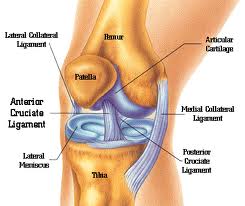
The knee is a joint that is made up of three parts. This joint has an inner (medial) and an outer (lateral) compartment. The kneecap (patella) joins the femur to form a third compartment called the patello femoral joint. The thighbone (femur) meets the large shinbone (tibia), forming the main knee joint.
The knee joint is covered with a joint capsule with ligaments strapping the inner and outer of the joint (collateral ligaments) as well as crossing within the joint (cruciate ligaments). These ligaments helps the knee to give stability and strength.

The meniscus is a thickened cartilage pad between the two joints formed by the femur and tibia. The meniscus acts as a smooth surface for motion and absorbs the load of the body above the knee when standing. The knee joint is covered by fluid-filled sacs called bursae, which act as gliding surfaces that reduce friction of the tendons. Below the kneecap, there is a large tendon called(patellar tendon) which attaches to the front of the tibia bone. Large blood vessels passing through the area behind the knee is called the popliteal space. The large muscles of the thigh move the knee. In the front of the thigh, the quadriceps muscles extend the knee joint. In the back of the thigh, the hamstring muscles flex the knee. The knee also rotates slightly under guidance of specific muscles of the thigh

Functions of the knee

The knee functions to allow movement of the leg and is critical to normal . Walking.The knee flexes normally to a maximum of 135 degrees and extends to 0 degrees. The bursae, or fluid-filled sacs, serve as gliding surfaces for the tendons to reduce the force of friction as these tendons move. The knee is a weight-bearing joint. Each meniscus serves to evenly load the surface during weight-bearing and also aids in disbursing joint fluid for joint lubrication.
The main function of the knee is to bend and straighten for moving the body. It also twists and rotates. Bones, ligaments, tendons, and cartilage.are the structures to help the knee to perform all the acions .
    Bones
    • The knee joint involves three bones.
    • The thighbone or femur comprises the top portion of the joint.
    • One of the bones in the lower leg (calf area), the tibia, provides the bottom portion of the joint.
    • The remaining bone in the calf, the fibula is not directly involved in the knee joint.
Ligaments
    • Ligaments are fibrous bands that connect bones to each other.
    • The knee includes four important ligaments, all four of which connect the femur to the tibia:
        The anterior cruciate ligament (ACL) and posterior cruciate ligament (PCL) provide front and back (anterior and posterior) and rotational stability to the knee.

Tendens

    • Instead of connecting bones to other bones as ligaments do, tendons connect muscles to bones.
    • The two important tendons in the knee are (1) the quadriceps tendon connecting the quadriceps muscle, which lies on the front of the thigh, to the patella and (2) the patellar tendon connecting the patella to the tibia (technically this is a ligament because it connects two bones).
    • The quadriceps and patellar tendons as well as the patella sometimes called the extensor mechanism, and together with the quadriceps muscle they facilitate leg extension (straightening)
      Cartilage 
    • Cartilaginous structures callled (one is a meniscus) line the top of the tibia and lie between the tibia and the two( knuckles )at the bottom of the femur (the femoral condyles).
    • Meniscus provide both space and cushion for the knee joint.
  • Bursae

    • Bursae (singular is bursa) are fluid-filled sacs that help to cushion the knee. The knee contains three important groups of bursae.
      • The prepatellar bursa
      • The anserine bursa
      • The infrapatellar bursa